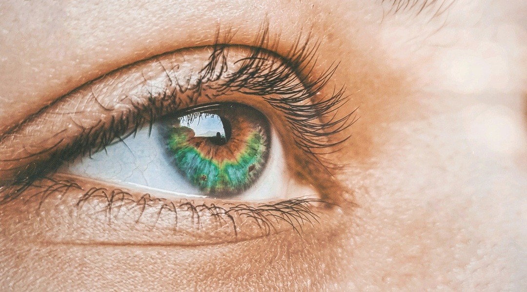
Retinal drug delivery is a mess.
It frustrates ophthalmologists because, for all our technological advancements, getting medicine to the back of the eyeball remains a logistical nightmare. Inject drugs directly into the vitreous? Half of them bounce off or get cleared out before they ever touch the retina. That is the main target. That is where the disease lives.
But researchers at Ghent University have figured out a way to actively steer medications to the back of the eye, and the results are promising enough to rethink how we treat blindness-causing conditions.
This isn’t just theoretical. We are talking about precise drug delivery to treat specific, devastating diseases like guided nanoparticles drug delivery for retinal treatment.
Age-related macular degeneration. Diabetic retinopathy. These conditions demand precision. If you miss the target, the treatment fails. Or worse, the side effects outweigh the benefits.
The Jelly Problem
Why is this so hard? It comes down to anatomy.
Between your lens and your retina sits the vitreous humor. It is a gel-like network. Collagen. Biomolecules. Its job is to hold your eye’s shape. It is dense. It is sticky. It is an obstacle course for drug molecules.
“Diffusion is slow. Heterogeneous. Hard to predict,” says Léa Guerassimoff, postdoctoral researcher at Ghent University’s Laboratory of General Biochemistry and Physical Pharmacy.
You inject a drug. It sits there. It drifts. By the time it reaches the retina, the concentration might be too low to work. So doctors do what they’ve always done: inject again. And again. And again.
More injections mean higher risks. Infection. Retinal detachment. Inflammation. It is a vicious cycle.
Steering the Particles
The solution? Stop fighting the gel and start riding it.
The team combined nanoparticles—tiny carriers designed to hold drugs—with indocyanine green. This is no exotic, untested chemical. It is a dye ophthalmologists use every day to visualize eye tissue during surgery. Familiar. Safe. Proven.
Here is the trick:
They pulse a laser into the vitreous. Short bursts. Nanoseconds.
The dye absorbs that energy. It heats up. Locally. Intensely.
That heat creates a physical reaction. Not the kind you’d expect.
A Surprise in the Physics
The researchers assumed they were using thermophoresis. That’s the force that pushes individual particles up or down a temperature gradient. It sounds logical. It makes sense on paper.
It turns out they were wrong.
The real mechanism is thermal convection.
When the laser hits the dye, the heated vitreous starts moving. Tiny, localized currents circulate. They swirl. And the nanoparticles? They just hitch a ride.
Without the dye, the particles—polystyrene in this test, uncharged—just drifted randomly. Useless.
With the laser and the dye, the drug cargo moved exactly where the team wanted it to go.
“This is the first time directed transport of drug cargo in the vitreous humor has been demonstrated,” Guerassimoff states.
Tuning the Treatment
It’s not magic. It’s physics you can adjust.
Want stronger steering? Crank up the dye concentration. Or increase the laser intensity.
Worried about particle size? Larger particles struggle to move through the gel. Smaller ones glide easier. The system responds.
But biology gets complicated.
The vitreous changes as you age. It liquefies. It forms pockets. Liquid pockets behave differently than solid gel. Particles move faster. They react differently to the laser.
This matters.
If your vitreous is liquefied due to aging, the treatment parameters change. One size does not fit all.
“This highlights the need for personalized approaches,” Guerassimoff notes. Patient-specific variations in the vitreous state must be considered in routine clinical practice.
From Lab to Clinic?
The potential is real.
Imagine a future where you get an injection. This one carries the drug, loaded onto nanoparticles, plus the dye. Then, the doctor applies a targeted laser pulse.
The laser guides the medication straight to the retina.
Higher drug localization. Better efficacy. Fewer repeat injections. Less risk of complications.
But hold your horses.
This is still proof of concept.
The experiments were done in controlled settings. The vitreous came from bovine eyes. Cow eyes. Not human eyes.
Safety studies are next. Human trials are far off.
Yet, there is a reason for cautious optimism.
They are using approved dyes. Clinically relevant laser wavelengths. The tools are already in the hospital drawer. We just need to learn how to use them together.
That alignment with existing practice could accelerate adoption. If the infrastructure is already there, scaling up is faster than building from scratch.
The eye remains a barrier. The vitreous is a wall. But walls have doors, if you know how to open them.